More Related Content
Similar to Chapt23 pregnancy & growth
Similar to Chapt23 pregnancy & growth (20)
Chapt23 pregnancy & growth
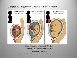
- 1. South Arkansas Community College Edited by B. Holmes MSN/Ed, RN Associate Professor Copyright © The McGraw-Hill Companies, Inc. Permission required for reproduction or display Chapter 23 Pregnancy, Growth & Development
- 2. Hole’s Human Anatomy and Physiology Twelfth Edition Shier Butler Lewis Chapter 23 Pregnancy, Growth, And Development Copyright © The McGraw-Hill Companies, Inc. Permission required for reproduction or display.
- 10. Copyright © The McGraw-Hill Companies, Inc. Permission required for reproduction or display. Zona pellucida Zygote Day 0 Ovulation Uterus Endometrium Stem cells Cleavages (first cleavage completed about 30 hours after fertilization) Stem cells Sperm nucleus Egg nucleus Polar bodies Day 4 Late morula Day 3 Early morula Day 2 4-cell stage Day 1 2-cell stage Pronucleus formation begins First cleavage division Fertilization occurs about 12-24 hours after ovulation Day 6-7 Blastocyst implantation
- 11. Copyright © The McGraw-Hill Companies, Inc. Permission required for reproduction or display Blastocyst (a) Trophoblast Inner cell mass Uterine wall (b) Invading trophoblast c: Courtesy of Ronan O'Rahilly, M.D. Carnegie Institute of Washington (c) Inner cell mass Endometrium Trophoblast Copyright © The McGraw-Hill Companies, Inc. Permission required for reproduction or display. Courtesy of Ronan O'Rahilly, M.D. Carnegie Institute of Washington Lumen Endometrium
- 14. Copyright © The McGraw-Hill Companies, Inc. Permission required for reproduction or display. Trophoblast cells secrete hCG hCG maintains corpus luteum Corpus luteum continues to secrete estrogens and progesterone Estrogens and progesterone promote growth, development, and maintenance of uterine wall
- 15. Copyright © The McGraw-Hill Companies, Inc. Permission required for reproduction or display. 0 2 4 Months of pregnancy 1 3 5 7 9 6 8 Increasing hormone concentration Estrogens Progesterone Human chorionic gonadotropin
- 20. Copyright © The McGraw-Hill Companies, Inc. Permission required for reproduction or display. Yolk sac Ectoderm Mesoderm Connecting stalk Skin Brain Chorion Heart Amnion Neural tube (Spinal cord) Amniotic fluid Digestive tract Chorionic villi Tail end Allantois Endoderm
- 21. Copyright © The McGraw-Hill Companies, Inc. Permission required for reproduction or display. a,b: © 2007 Landrum B. Shettles; c: © Petit Format/Nestle/Photo Researchers, Inc. (a) (b) (c)
- 22. Copyright © The McGraw-Hill Companies, Inc. Permission required for reproduction or display. b: © Carroll Weiss/Camera M.D. Studios (b) Actual length 4 weeks 5 weeks 6 weeks 7 weeks (a) Actual length Actual length Actual length
- 23. Copyright © The McGraw-Hill Companies, Inc. Permission required for reproduction or display. – (e) 49 ± 1 day (28–30 mm) Developing ear External ear Forebrain Elbow Handplate Lens Midbrain Ear Eyelid Heart prominence Hindlimb External ear Digital rays Pigmented eye Paddle-shaped foot plate Notches between toe rays Webbed fingers Mandibular process Paddle-shaped forelimb External acoustic meatus Fingers separated Toes separated Fan-shaped webbed toes (c) 40 ± 1 day (16–21 mm) (a) 35 ± 1 day (10–12 mm) (b) 37 ± 1 day (12.5–15.75 mm) (d) 45 ± 1 day (22–24 mm) (g) 56 ± 1 day (34–40 mm) (f) 52 ± 1 day (32–34 mm) Maxillary process Developing eye Toe rays Wrist
- 25. Copyright © The McGraw-Hill Companies, Inc. Permission required for reproduction or display. Artery Chorion Chorionic villi Connective tissue Section of villus Lacuna filled with maternal blood Placental membrane Wall of villus Maternal blood Embryonic capillaries Vein Copyright © The McGraw-Hill Companies, Inc. Permission required for reproduction or display. Umbilical cord Umbilical arteries Umbilical vein Placenta Endometrium Lacuna Chorion Myometrium Decidua basalis (maternal portion of placenta) Maternal blood vessels Embryonic blood vessels Villi (embryonic portion of placenta)
- 26. Copyright © The McGraw-Hill Companies, Inc. Permission required for reproduction or display. Chorion Placenta Umbilical cord Amniochorionic membrane Endometrium Myometrium Amniotic fluid
- 27. Copyright © The McGraw-Hill Companies, Inc. Permission required for reproduction or display. © Donald Yaeger/Camera M.D. Studios Copyright © The McGraw-Hill Companies, Inc. Permission required for reproduction or display. 0 Accutane 1 2 3 4 5 6 7 8 9 Month (b) When different teratogens disrupt development 0 1 2 3 4 5 6 7 8 9 Eyes Ears Heart Month (a) When physical structures develop Reproductive system Upper and lower limbs Central nervous system Diethylstilbestrol Thalidomide
- 29. Copyright © The McGraw-Hill Companies, Inc. Permission required for reproduction or display. 13 years 2 month embryo 3 month fetus 22 years Newborn 2 years 5 years
- 30. Copyright © The McGraw-Hill Companies, Inc. Permission required for reproduction or display. Genital tubercle Urogenital folds Labioscrotal folds Fused urogenital folds Perineum Anus Urethral groove (a) Urogenital fold Developing penis (c) (e) (d) (f) Glans penis Scrotum Genital tubercle Glans Urogenital fold Labioscrotal fold (b) Developing clitoris Embryonic tail Urethral groove Glans clitoris Hymen Labia minora Labia majora Perineum Anus Urethral orifice Prepuce Male Female Vaginal orifice
- 31. Copyright © The McGraw-Hill Companies, Inc. Permission required for reproduction or display. Umbilical cord Placenta Uterine wall Cervix Amniochorionic membrane Amniotic fluid
- 34. Fetal Blood and Circulation Copyright © The McGraw-Hill Companies, Inc. Permission required for reproduction or display. Placenta Fetal capillaries Umbilical vein Umbilical arteries Uterine wall Maternal blood in lacuna Diffusion Oxygen and nutrients into fetal blood Diffusion W aste substances into maternal blood Chorionic villus Blood flow from fetus, branch of umbilical artery Blood flow to fetus, branch of umbilical vein
- 35. Copyright © The McGraw-Hill Companies, Inc. Permission required for reproduction or display. Aortic arch Superior vena cava Inferior vena cava Hepatic portal vein Pulmonary artery Left atrium Pulmonary veins Abdominal aorta Pulmonary trunk Left ventricle Left renal artery Common iliac artery Internal iliac artery Umbilical vein Umbilical arteries Placenta Foramen ovale (becomes fossa ovalis) Ductus venosus (becomes ligamentum venosum) Umbilical vein (becomes ligamentum teres) Umbilical arteries (become medial umbilical ligaments) Ductus arteriosus (becomes ligamentum arteriosum) Decreasing blood oxygen level
- 36. Copyright © The McGraw-Hill Companies, Inc. Permission required for reproduction or display. Placenta Liver Lungs Ductus venosus Right atrium Right ventricle Foramen ovale Left atrium Left ventricle Aortic arch Aorta Umbilical vein (oxygen, nutrients) Superior vena cava Inferior vena cava Heart, brain, upper limbs Ductus arteriosus (most of the blood) Pulmonary trunk Trunk and lower limbs Umbilical artery (carbon dioxide, wastes) Umbilical artery (carbon dioxide, wastes) Internal iliac arteries Decreasing blood oxygen level
- 40. Copyright © The McGraw-Hill Companies, Inc. Permission required for reproduction or display. Fetal head is forced toward cervix Cervix is stretched Stretch receptors are stimulated Fetus is moved downward Reflex is elicited that causes stronger uterine contractions
- 41. Copyright © The McGraw-Hill Companies, Inc. Permission required for reproduction or display. Placenta Urethra V agina Cervix Rectum Amniotic sac (b) (a) (c) (d) Placenta Placenta Uterus Symphysis pubis Urinary bladder Ruptured amniotic sac Umbilical cord
- 43. © Biophoto Associates/Photo Researchers, Inc. (a) (b) Glandular tissue with secretions Glandular tissue Connective tissue Copyright © The McGraw-Hill Companies, Inc. Permission required for reproduction or display.
- 44. Copyright © The McGraw-Hill Companies, Inc. Permission required for reproduction or display. Release Duct Lumen Myoepithelial cells Secretion Milk Nipple or areola of breast is stimulated Nerve impulses travel to hypothalamus Hypothalamus signals posterior lobe of pituitary gland to release oxytocin Oxytocin causes myoepithelial cells surrounding alveolar glands to contract Milk is released from ductile system through nipple Copyright © The McGraw-Hill Companies, Inc. Permission required for reproduction or display.